Case Presentation
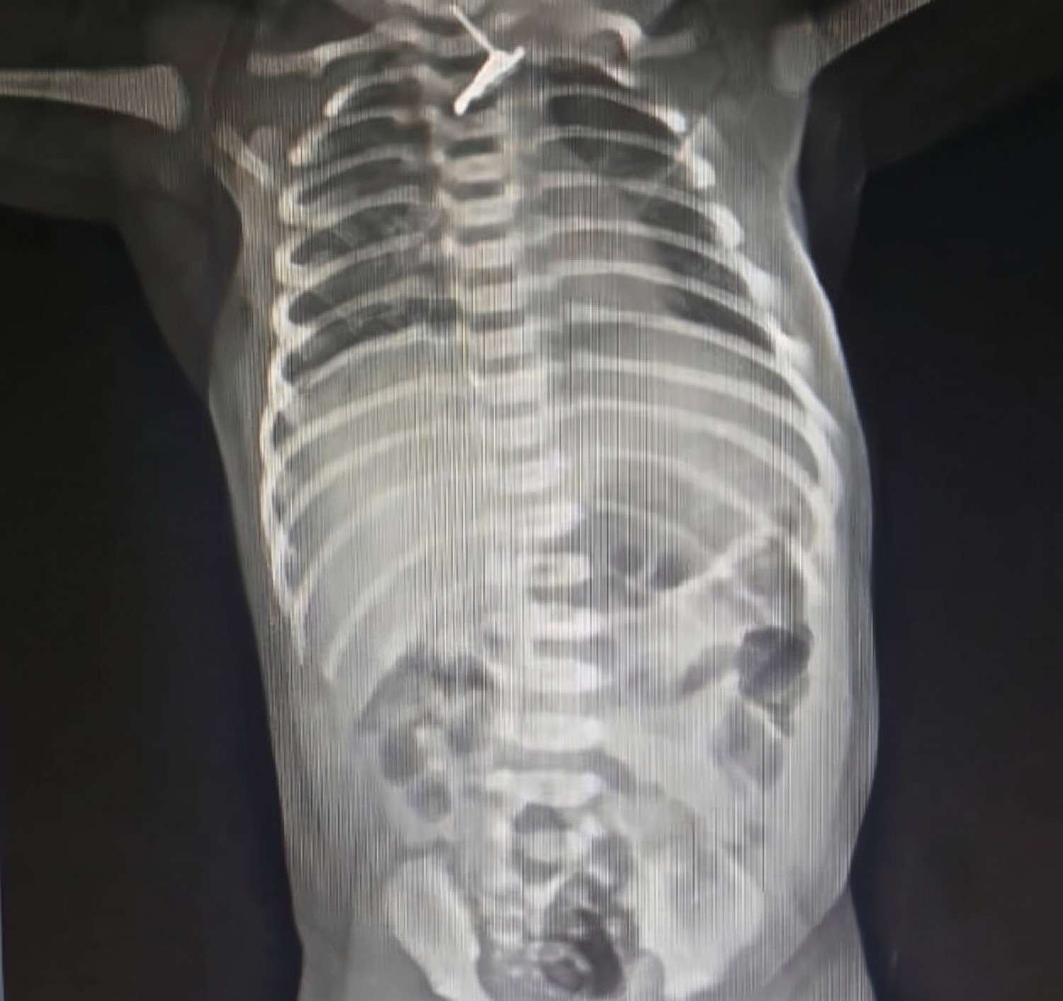
A 5-day-old term male neonate weighing 3.4 kg was brought to our hospital following accidental insertion of an ear ring into his mouth by his 2-year-old sibling. The antenatal, birth and immediate neonatal periods had been uneventful.
The incident had occurred approximately 4 hours prior to presentation. On arrival, the neonate was clinically stable, with no respiratory distress, stridor, cyanosis or other evidence of airway compromise. He was able to breastfeed, although a minimal episode of vomiting was noted.
Initial Assessment
In view of the suspected upper aerodigestive tract foreign body, the neonate was kept nil per oral and observed while arrangements were made for endoscopic evaluation.
After approximately 4 hours, he was taken to the operating theatre for controlled endoscopic foreign-body retrieval.
 Enlarge
Endoscopic Retrieval
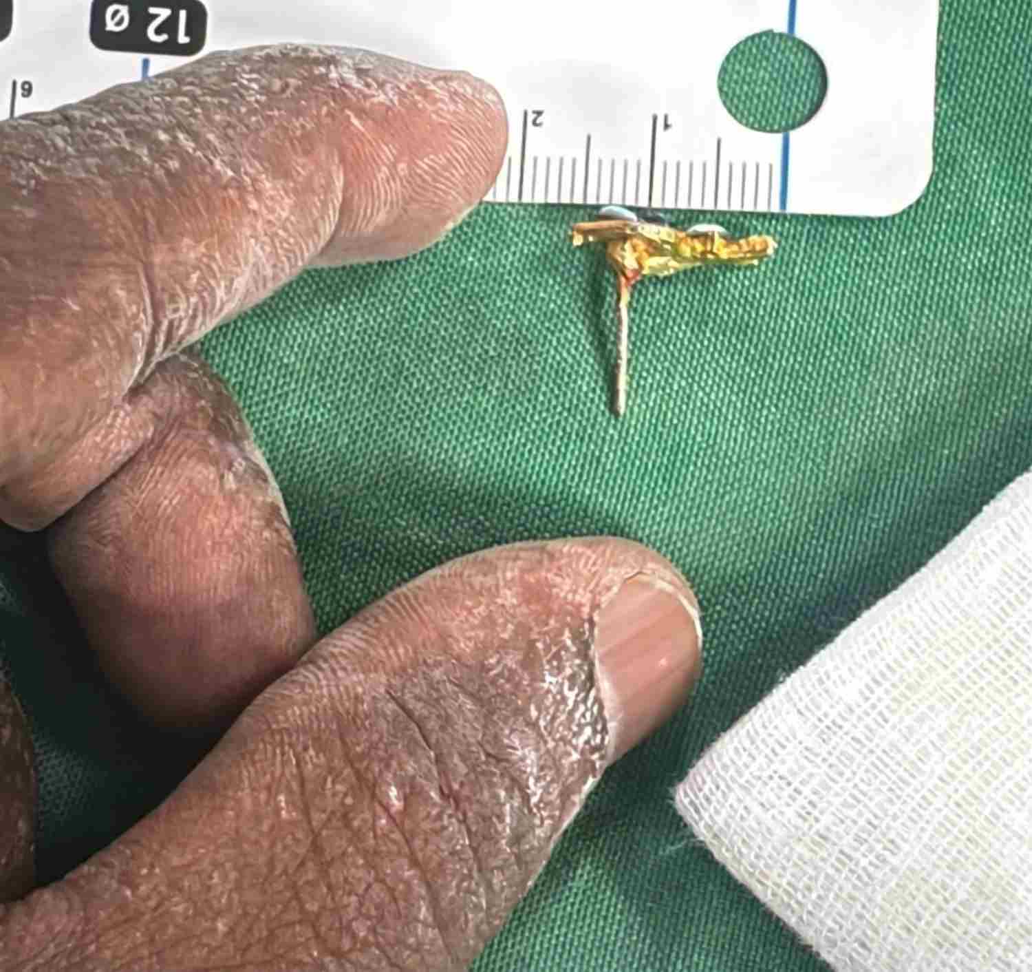
Endoscopic examination demonstrated an ear ring impacted at the level of the cricopharynx.
The foreign body was found to be partially embedded in the mucosa. It was carefully mobilized and retrieved under direct visualization, taking particular care to avoid further mucosal trauma.
Following removal, only minimal mucosal bleeding was noted. The upper aerodigestive tract was inspected and there was no obvious large mucosal defect identified.
 Enlarge
Post-Procedure Complication
The neonate was immediately extubated following the procedure and placed on supplemental oxygen through nasal cannula. He was transferred to the neonatal intensive care unit for close monitoring.
During the post-procedure period, the neonate developed subcutaneous emphysema and pneumomediastinum.
- Subcutaneous emphysema
- Pneumomediastinum
In view of the stable clinical condition and absence of respiratory compromise, conservative management was adopted.
Conservative Management
The neonate was maintained nil per oral for 72 hours and received supportive and anti-edema measures along with supplemental nasal oxygen.
The subcutaneous emphysema and pneumomediastinum progressively resolved over the subsequent 24–48 hours without the need for surgical intervention or chest drainage.
-
0 Hours
Procedure
-
Post-procedure
Monitoring
-
24–48 Hours
Subcutaneous emphysema and pneumomediastinum progressively resolved
-
72 Hours
Feeds cautiously reintroduced
-
Day 5
Full feeds achieved
Recovery
After 72 hours, feeds were cautiously reintroduced following clinical assessment for evidence of an ongoing aerodigestive tract leak.
Feeding was gradually advanced and was well tolerated.
The neonate reached full feeds by post-procedure day 5 and remained clinically stable without respiratory or feeding-related complications.
- Full feeds achieved by post-procedure day 5
- No respiratory complications
- No feeding-related complications
- No surgical intervention required
Why This Case Matters
Three unusual aspects made this case clinically distinctive.
An exceptionally young patient
A foreign body in the upper aerodigestive tract of a 5-day-old neonate is exceptionally unusual.
An uncommon site of impaction
An ear ring became impacted in the cricopharyngeal mucosa, requiring careful endoscopic extraction.
A complication managed without surgery
The development of pneumomediastinum and subcutaneous emphysema was managed conservatively, with resolution without surgical exploration or chest drainage.
Even when a neonate presents without obvious respiratory compromise, an upper aerodigestive foreign body requires careful evaluation and a controlled approach to airway protection and retrieval. In this case, close monitoring after endoscopic removal allowed post-procedure complications to be managed conservatively, with gradual return to feeding and an uncomplicated recovery.